


What Is an Elastic Bandage and When Should It Be Used?
Views: 0 Author: Site Editor Publish Time: 2026-08-24 Origin: Site


Compression therapy manages acute injuries and post-operative recovery by controlling localized swelling and stabilizing damaged tissues. Using the wrong wrap leads to poor edema control, inadequate joint support, or severe complications like tissue ischemia. We see this often in field trauma and clinical wards where improper application causes a dangerous tourniquet effect. You cannot just grab any wrap from a first-aid kit and expect optimal results. This guide provides a technical evaluation framework for medical staff, athletic trainers, and procurement teams. We will cover how to select, evaluate, and apply the right Elastic Bandage for specific medical and sports applications. You will learn stretch dynamics, closure mechanisms, and application protocols to ensure patient safety and functional recovery. Stop relying on outdated materials and start implementing evidence-based compression strategies to improve patient outcomes.
Key Takeaways
Primary Function: An elastic compression bandage is designed to provide localized, even pressure to reduce swelling, decrease pain, and stabilize minor musculoskeletal injuries, not to immobilize fractures.
Material Matters: Selecting between latex, latex-free, short-stretch, and long-stretch variations is critical for patient safety and specific clinical outcomes (e.g., sports injuries vs. lymphedema).
Closure Mechanisms Dictate Usability: Hook-and-loop (Velcro) closures offer superior reliability and safety compared to traditional metal clips, which pose skin tear and ingestion risks.
Application Risk: Improper wrapping techniques can restrict arterial blood flow; standardized application protocols (like the figure-eight method) and capillary refill monitoring are mandatory for safe use.
What Is an Elastic Bandage? (Defining the Baseline)
To utilize compression effectively, you must understand the physical mechanics of the tools involved. The baseline definition sets the standard for clinical expectations and application methods across different medical disciplines.
Mechanical Definition
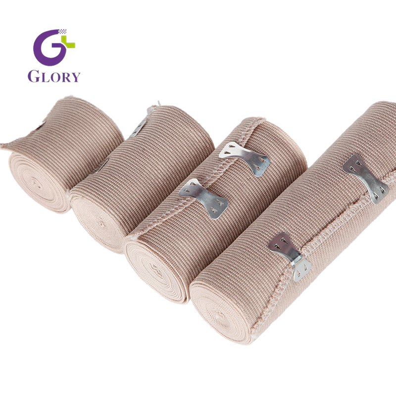
An elastic bandage is a stretchable, woven, or knitted fabric strip designed to conform closely to body contours. Its primary mechanical function is to exert consistent, graded pressure across a specific anatomical area. This pressure manages localized hemodynamics and acute inflammation. The fabric incorporates elastic fibers—traditionally rubber, but increasingly synthetic elastomers—that allow the material to stretch under tension and return to its original length. This memory effect maintains continuous compression against the skin. It adapts to changes in swelling and muscle movement without losing its structural integrity or requiring constant readjustment.
Clinical Objectives
In acute injury management, this tool forms the foundation of the R.I.C.E. (Rest, Ice, Compression, Elevation) protocol. External mechanical pressure limits interstitial fluid accumulation, commonly known as edema. By compressing the tissue, the bandage forces excess fluid out of the interstitial spaces and back into the lymphatic and venous systems for clearance. Furthermore, this compression reduces pain by relieving tissue tension on localized nerve endings. Beyond fluid management, the wrap provides a layer of physical protection and enhances proprioceptive feedback. This heightened spatial awareness helps patients avoid moving injured joints into painful or damaging ranges of motion during the early stages of recovery.
Terminology Clarification
Medical terminology often blurs with commercial branding, creating confusion during procurement. Differentiate the generic medical device from proprietary brand names like Tensor bandage or ACE wrap, which have become colloquialisms for the product category. In professional and clinical literature, the term is entirely interchangeable with Elastic Compression Bandage. Regardless of the brand name printed on the packaging, the underlying mechanical function, stretch dynamics, and clinical evaluation criteria remain identical. Focus on the material specifications rather than the marketing label.
When Should an Elastic Bandage Be Used? (Clinical Success Criteria)
Applying compression is not a universal remedy for all musculoskeletal trauma. Identifying the correct clinical indications ensures efficacy, while recognizing contraindications prevents severe iatrogenic injury.
Appropriate Indications (Target Use Cases)
Targeted use cases maximize the benefits of graded compression. The following scenarios represent the standard indications for these wraps in clinical and field settings.
Acute Sprains and Strains: The most common application involves managing initial swelling and decreasing localized pain following minor muscle and ligament injuries, such as a Grade 1 ankle sprain. The wrap provides mild kinematic support without completely restricting mobility.
Post-Operative Edema Management: Following orthopedic procedures like ACL reconstructions or joint replacements, surgeons utilize controlled compression to prevent fluid pooling in the lower extremities. This proactive management reduces the risk of deep vein thrombosis (DVT) and accelerates tissue healing.
Sports Medicine and Prophylaxis: Athletes frequently use proactive wrapping for joint support and physical protection during high-impact activities. This proves particularly useful when rigid taped alternatives are unavailable, inappropriate, or too restrictive for the required range of motion on the field.
Securing Dressings: These wraps act as an excellent secondary dressing. They hold primary wound care materials, sterile gauze, or cold packs firmly in place. This method avoids the adhesive skin damage often caused by medical tapes, especially on fragile, pediatric, or aging skin.
Contraindications (When NOT to Use)
Improper application can exacerbate existing medical conditions. Recognize these strict contraindications before applying any circumferential pressure to a patient.
Arterial Insufficiency: Applying compression to patients with peripheral arterial disease (PAD) or severely compromised circulation is highly dangerous. Even mild external pressure can collapse weakened arteries, leading to rapid tissue ischemia, necrosis, and potential amputation.
Acute Fractures or Severe Instability: Flexible wraps do not provide the rigid immobilization required for broken bones, severe joint dislocations, or complete ligament ruptures. Relying on flexible fabric for unstable fractures risks further bone displacement and internal tissue damage.
Overnight Wear: Leaving compression wraps on during sleep introduces significant risk. Swelling fluctuates overnight, and the lack of conscious sensory monitoring means a patient will not feel the warning signs of restricted blood flow. This leads to localized nerve damage or acute compartment syndrome.
Pre-Application Assessment Protocol
Before applying any compression therapy, clinicians and first responders must evaluate the patient to rule out contraindications. Follow this standard assessment sequence:
Verify the injury mechanism to rule out acute fractures or complete structural failures.
Assess baseline circulation by checking distal pulses and capillary refill times.
Confirm the patient has no history of peripheral arterial disease or severe neuropathy.
Select the appropriate wrap width based on the specific anatomical site being treated.
Types of Elastic Compression Bandages (Solution Categories)
Not all stretchable fabrics perform the same function. The specific construction, stretch dynamics, and material formulation dictate the clinical application and the resulting patient outcomes. Selecting the wrong type compromises the entire treatment plan.
Short-Stretch vs. Long-Stretch Dynamics
The most critical mechanical distinction lies in the stretch capacity and the resulting pressure dynamics. Long-stretch bandages possess high elasticity, often stretching up to 140% or more of their original length. They deliver high resting pressure, meaning they continuously squeeze the limb even when the patient is completely still. This continuous pressure makes them best suited for acute sports injuries, temporary joint support, and active movement scenarios where immediate edema control is required.
Conversely, short-stretch bandages contain inextensible fibers that limit their stretch to roughly 30-60%. They exert low resting pressure but high working pressure. When a patient moves, their muscles expand against the rigid barrier of the short-stretch fabric. This creates a mechanical pumping action that forces lymphatic fluid upward against gravity. This dynamic makes short-stretch variants ideal for managing chronic edema, lymphedema, and venous leg ulcers over extended periods.
Woven vs. Knitted Construction
Manufacturing techniques directly impact durability, breathability, and cost. Woven bandages feature a tighter, interlocking thread pattern. This construction is highly durable, offers heavier compression, and maintains its structural integrity through repeated stretching and washing. However, the dense weave can trap heat against the skin, causing discomfort in warm environments.
Knitted bandages are constructed with looped threads, resulting in a lighter, more breathable fabric. They conform easily to complex anatomical shapes like elbows and ankles, and they cost significantly less to manufacture. The trade-off is long-term durability. Knitted variants degrade, lose their elasticity, and unravel faster under heavy clinical use or frequent laundering.
Latex vs. Latex-Free Formulations
Historically, natural rubber latex provided the necessary elasticity for these wraps. However, the medical industry has experienced a massive shift toward 100% latex-free synthetic elastomers. This transition eliminates the risk of severe anaphylactic reactions in both patients and healthcare staff. Modern latex-free formulations, utilizing materials like spandex or elastane, now match or exceed the stretch memory and durability of traditional rubber. Latex-containing wraps are largely obsolete and represent an unnecessary liability in modern clinical procurement.
Comparison of Stretch Dynamics
Feature | Long-Stretch | Short-Stretch |
|---|---|---|
Stretch Capacity | Up to 140%+ | 30% - 60% |
Resting Pressure | High | Low |
Working Pressure | Low to Moderate | High |
Primary Clinical Use | Acute sprains, sports injuries, joint support | Lymphedema, chronic edema, venous ulcers |
Wear Time | Short-term (remove at night) | Long-term (can often be worn overnight) |
Evaluation Dimensions: How to Choose the Right Elastic Bandage
Selecting the correct product requires matching the physical specifications of the wrap to the anatomical requirements of the patient. Evaluate sizing, closure methods, and lifecycle costs to ensure optimal results and prevent application errors.
Sizing and Width Selection (Anatomy-Based Matching)
Applying the wrong width compromises both compression gradients and joint mobility. A wrap that is too narrow will cut into the skin, acting like a tourniquet and restricting blood flow. A wrap that is too wide will bunch up behind joints, causing severe friction blisters and uneven pressure distribution.
Sizing Guide by Anatomical Region
Bandage Width | Target Anatomy | Clinical Application Notes |
|---|---|---|
2-Inch | Fingers, wrists, hands, pediatric limbs | Ideal for small radius curves; prevents bulky overlap on small joints. |
3-Inch | Adult ankles, forearms, elbows | The standard size for most common sports injuries and upper extremity trauma. |
4-Inch | Lower legs, large ankles, small knees | Provides excellent coverage for calf strains and robust ankle support. |
6-Inch | Knees, thighs, shoulders, chest | Required for massive muscle groups to ensure even pressure distribution. |
Closure Mechanisms (Feature-to-Outcome Evaluation)
The method used to secure the wrap dictates its safety and reliability during movement. Traditional metal clips feature sharp prongs that hook into the fabric. These are increasingly obsolete due to high loss rates, skin puncture risks, and a complete lack of MRI compatibility. Metal clips also cause severe injury if accidentally ingested by pediatric or veterinary patients.
Hook-and-loop (Velcro) fasteners represent the modern standard. They provide secure, self-contained fastening that maintains consistent tension and prevents unravelling during high-intensity activity. Because the fastening mechanism is built directly into the ends of the fabric, there are no loose parts to lose in a busy trauma bay or athletic training room.
Self-adhering (cohesive) wraps offer a hybrid solution. These materials are coated with a cohesive substance that sticks firmly to itself but not to skin, hair, or underlying dressings. Cohesive wraps are ideal for veterinary use, pediatric care, or highly active human applications where frequent readjustment is necessary and adhesive tape is contraindicated.
Durability, Washability, and Scalability
Evaluate the cost-per-use economics when stocking a clinic or athletic training room. Cheap, single-use wraps lose their elasticity rapidly and contribute heavily to medical waste. Premium, washable bandages retain their stretch memory through multiple laundering cycles, offering better long-term value and consistent clinical performance.
Maintaining this elasticity requires strict adherence to care instructions. Wash the fabric by hand using lukewarm water and a mild detergent. Never use bleach or fabric softeners, as these harsh chemicals destroy synthetic elastomers. After washing, lay the fabric flat on a clean towel to air dry. Hanging the wet fabric vertically causes the weight of the water to stretch and permanently ruin the elastic fibers.
Assessing Material Degradation
Knowing when to discard a wrap is just as important as knowing how to apply it. Using degraded materials results in zero compression and false security. Inspect your inventory regularly for the following signs of failure:
Scalloped or frayed edges that indicate the structural weave is breaking down.
A complete loss of recoil, where the fabric remains stretched out after removal.
Thinning patches in the material that create uneven pressure zones.
Velcro fasteners that no longer grip securely, risking sudden unravelling during patient movement.
Implementation Risks and Application Protocols
Even the highest quality material becomes dangerous if applied incorrectly. Standardized application protocols mitigate risks and ensure the compression serves its intended clinical purpose without causing secondary injuries.
Mitigating the Tourniquet Effect
The most severe risk of improper application is the tourniquet effect. If wrapped too tightly, the fabric occludes venous return before it occludes arterial supply. Blood pumps into the limb but cannot escape, leading to massive distal swelling, severe throbbing pain, and eventual tissue ischemia.
To prevent this, follow the 50/50 rule: apply the fabric with 50% stretch and overlap each previous layer by exactly 50%. Always begin wrapping at the most distal point (farthest from the heart) and move proximally. This creates a graded compression effect—tighter at the bottom, looser at the top—which physically pushes fluid back toward the body's core.
Monitoring circulation post-application is mandatory. Check capillary refill by pressing on the patient's nail bed; the pink color should return within two seconds. Monitor skin temperature to ensure the extremity does not become cold. Finally, ask the patient for sensory feedback. Any reports of numbness, tingling, or throbbing indicate the wrap is too tight and must be removed immediately.
The Figure-Eight Wrapping Technique
For joint applications, the figure-eight method is biomechanically superior to standard spiral wrapping. Spiral wrapping around an ankle or wrist tends to bunch up in the crease of the joint during movement, causing severe friction, restricted blood flow, and uneven pressure.
The figure-eight technique creates a structural application that prevents bunching, ensures even pressure distribution, and maximizes joint protection. By crossing the joint line diagonally, the fabric creates a mechanical block against excessive inversion or eversion while still allowing necessary flexion and extension.
Follow these steps for a proper figure-eight application on an ankle:
Anchor the fabric with two straight, overlapping turns around the mid-foot, just behind the toes.
Pull the fabric diagonally upward across the anterior joint line (top of the foot) toward the lower leg.
Wrap completely around the lower leg, just above the ankle bones (malleoli).
Cross back down diagonally over the top of the foot to form an "eight" pattern.
Repeat this figure-eight pattern, overlapping each previous layer by 50%, until the joint is fully supported.
Secure the end of the wrap with the hook-and-loop fastener on the lower leg, never directly over the joint crease.
Patient Education Post-Application
Clinical responsibility does not end once the wrap is secured. Patients must understand how to manage their compression therapy at home to prevent complications. Provide clear, actionable instructions before discharging the patient.
Instruct the patient to elevate the injured limb above heart level whenever possible to assist fluid drainage.
Warn them never to re-wrap the injury tighter than the clinician originally applied it.
Mandate the removal of the wrap before sleeping to prevent overnight compartment syndrome.
Teach them to recognize the signs of restricted circulation, including blue toes, cold skin, or a "pins and needles" sensation.
Conclusion
An elastic bandage remains a highly effective, low-cost intervention for managing edema, reducing localized pain, and stabilizing minor musculoskeletal trauma. However, this efficacy relies entirely on selecting the correct width, stretch-type, and closure mechanism for the specific anatomical target. Moving away from outdated materials and adopting modern, standardized application techniques prevents complications and accelerates recovery.
For general first-aid and sports medicine applications, long-stretch, hook-and-loop, latex-free bandages provide the best combination of safety, durability, and ease of use. Short-stretch variants should remain reserved for specialized vascular and lymphatic care under professional supervision.
Take the following steps to optimize your compression therapy protocols:
Audit your current clinical inventory and discard any degraded wraps that have lost their stretch memory or feature frayed edges.
Eliminate all bandages utilizing metal clips to completely remove puncture hazards and foreign-body ingestion risks.
Standardize your supply chain by sourcing exclusively latex-free materials to protect against severe allergic reactions.
Train all staff and patients on the 50/50 stretch-and-overlap rule and the biomechanically superior figure-eight wrapping technique.
FAQ
Q: How tight should an elastic compression bandage be?
A: It should feel snug and supportive but never restrictive or painful. Apply it using 50% of its maximum stretch capacity. You should be able to comfortably slide one or two fingers under the fabric. If the patient experiences throbbing, numbness, tingling, or cold toes, the wrap is too tight and must be completely removed and reapplied with less tension.
Q: Can you sleep with an elastic bandage on?
A: No, you should never sleep with a standard long-stretch wrap on. Swelling naturally decreases when you lie down and elevate the limb. Leaving the wrap on overnight can severely restrict blood flow while you sleep. Because you are unconscious, you will not feel the warning signs of numbness, which can lead to permanent tissue or nerve damage.
Q: What is the difference between a cohesive bandage and an elastic bandage?
A: A traditional elastic wrap relies on stretch memory and external fasteners like Velcro to stay in place. A cohesive bandage contains a specific chemical coating that makes the fabric stick firmly to itself without adhering to skin, hair, or underlying gauze. Cohesive wraps are typically single-use, whereas traditional wraps are highly durable, washable, and reusable.
Q: Can you wash and reuse an elastic bandage?
A: Yes, most high-quality wraps are designed for multiple reuses. Wash them gently by hand in lukewarm water with mild soap. Never use bleach or fabric softener, as these destroy the synthetic elastomers. To dry, lay the fabric flat on a clean towel. Do not hang it vertically; the weight of the water will stretch and ruin the elastic fibers.
Q: How do I know if my compression bandage is wrapped too tightly?
A: Watch for distinct physical signs: the skin distal to the wrap turning pale or blue, the extremity feeling cold to the touch, or a capillary refill time taking longer than two seconds. Subjective signs reported by the patient include increased throbbing pain, numbness, or a distinct "pins and needles" sensation in the affected limb.
Q: What width of elastic bandage is best for an ankle sprain?
A: A 3-inch or 4-inch width is the standard and most effective size for an adult ankle sprain. A 2-inch wrap is too narrow and may cut into the skin, restricting blood flow. Conversely, a 6-inch wrap is too wide and will bunch up uncomfortably inside the crease of the ankle joint, causing severe friction blisters.
Q: Does an elastic bandage help heal a sprain faster?
A: It does not directly heal the torn ligaments, but it creates an optimal physiological environment for healing. By controlling excess swelling and providing mild structural support, it reduces pain and prevents further mechanical irritation. This allows the body's natural inflammatory and repair phases to proceed efficiently without being hindered by massive edema.
