


What Factors Matter When Selecting a Wound Dressing?
Views: 0 Author: Site Editor Publish Time: 2026-07-30 Origin: Site


Improper dressing selection directly correlates with delayed healing, increased infection rates, and escalated care costs. Managing dynamic wound environments requires balancing fluctuating exudate, bioburden, and tissue phases. Clinicians face an overwhelming market of traditional and advanced products. Matching the right product to the specific wound bed is a daily clinical challenge. You need a systematic, evidence-based evaluation framework. Selecting a Wound Dressing relies on wound bed characteristics, patient-specific variables, and the total cost of healing. The TIME framework provides a structured approach to evaluate tissue, infection, moisture, and edges. Understanding these factors ensures you apply the most effective interventions at the right time. We will explore various dressing categories and their clinical applications. You will also discover how to balance operational workflows with patient outcomes and mitigate common implementation risks.
Dressing selection must be dictated by the current phase of wound healing (necrotic, sloughy, granulating, or epithelializing) and requires continuous reassessment.
Exudate management is the primary driver of dressing choice; balancing moisture to prevent both desiccation and peri-wound maceration is critical to maintaining a optimal microenvironment.
Advanced wound dressing products often present a higher unit cost but can reduce the total cost of care by decreasing dressing change frequency, reducing nursing time, and lowering complication rates.
Patient quality of life (pain, mobility, odor management) and compliance must be weighted equally with clinical indications to ensure successful protocol adherence.
Defining Success Criteria: The TIME Framework for Clinical Evaluation
Tissue Assessment (T): Wound Bed Composition and Debridement Needs
Evaluating the wound bed composition dictates your initial intervention strategy. Necrotic tissue or eschar requires active removal to promote healing. You must select moisture-donating dressings to facilitate autolytic debridement. Enzymatic or mechanical debridement also relies on proper moisture balance to function effectively. When dealing with hard, dry eschar, applying a hydrogel covered by a moisture-retentive film softens the dead tissue over several days. This allows the body's endogenous enzymes to break down the necrotic material safely.
Sloughy tissue presents a different clinical challenge. It requires continuous absorption and selective debridement. Dressings must manage the liquid slough without drying out the underlying healthy cells. Granulating and epithelializing tissue needs physical protection. These fragile cells require thermal insulation and strict moisture maintenance to migrate across the wound surface. Applying aggressive adhesives or highly absorbent materials to a clean, granulating wound bed strips away new cellular growth and restarts the inflammatory phase.
Infection and Inflammation Control (I): Bioburden and Infection Status
Distinguishing between contaminated, colonized, critically colonized, and infected wounds dictates your antimicrobial strategy. All chronic wounds contain bacteria. Not all require antimicrobial intervention. Contaminated and colonized wounds generally heal well with standard moisture management and routine cleansing. Overusing topical antimicrobials on these wounds leads to cellular toxicity and delays granulation.
Critical colonization stalls the healing process. You will notice increased exudate, friable granulation tissue, and lingering odor. This status triggers the transition from inert dressings to active antimicrobial or barrier dressings. Frank infection demands immediate topical and systemic treatment to prevent tissue destruction and systemic complications. You must monitor for spreading erythema, localized heat, increased pain, and purulent drainage. When biofilm is suspected, physical debridement combined with a targeted antimicrobial dressing is required to disrupt the protective bacterial matrix.
Moisture Balance (M): Exudate Volume and Viscosity Assessment
Moisture balance remains the cornerstone of modern wound care. You must match the dressing absorption capacity to the current exudate levels. Wounds produce varying amounts of fluid ranging from none to heavy. An overly dry wound bed halts cellular migration. An overly wet environment causes tissue maceration and breakdown. You must evaluate both the volume and the consistency of the drainage during every dressing change.
Exudate viscosity impacts dressing performance. Thick, viscous exudate clogs the pores of certain dressings. This leads to fluid pooling beneath the dressing layer. Pooling increases the risk of bacterial proliferation and periwound damage. Thin, watery exudate wicks rapidly through standard materials, requiring dressings with high retention capacities under compression.
Exudate Level | Clinical Goal | Recommended Dressing Type | Change Frequency Indicator |
|---|---|---|---|
None / Dry | Donate moisture, rehydrate tissue | Hydrogels, Hydrocolloids | Dressing dries out or loses adhesion |
Low | Maintain moisture, protect tissue | Thin Films, Hydrocolloids | Fluid reaches dressing margins |
Moderate | Absorb excess, maintain balance | Foams, Alginates | 75% saturation or strike-through |
Heavy | Maximize absorption, protect edges | Superabsorbents, Thick Foams | Approaching secondary layer saturation |
Edge of Wound (E): Assessing Wound Margins and Epibole
The status of the wound perimeter indicates the overall healing trajectory. Stalled or rolled edges, known as epibole, prevent epithelial cells from migrating across the wound bed. Calloused edges often surround diabetic foot ulcers and require sharp debridement. If the edges are not attached to the wound bed, epithelialization cannot occur.
Your dressing choice must support an advancing wound edge. It should protect migrating epithelial cells at the margins from trauma and excess moisture. Proper edge management ensures the wound can contract and close efficiently. Applying a barrier film to the periwound skin prevents maceration, keeping the edges intact and capable of inward migration.
Anatomical Architecture: Wound Depth, Cavity, and Tunneling
Deep wounds, cavities, and tunneling require specific structural considerations. You must pack dead space to prevent premature surface closure. Surface closure over an empty cavity leads to abscess formation and severe complications. Packing materials must conform to the cavity walls without applying excessive pressure, which can cause localized ischemia.
Dressings used in deep cavities require high tensile strength. They must remain intact during removal. Leaving dressing fragments behind in a deep tract triggers foreign body reactions and chronic inflammation. Always count the pieces inserted and removed. Use single, continuous strips of packing material rather than multiple small pieces to ensure complete retrieval.
Evaluating Wound Dressing Products: Categories and Clinical Applications
Traditional and Contact-Layer Dressings
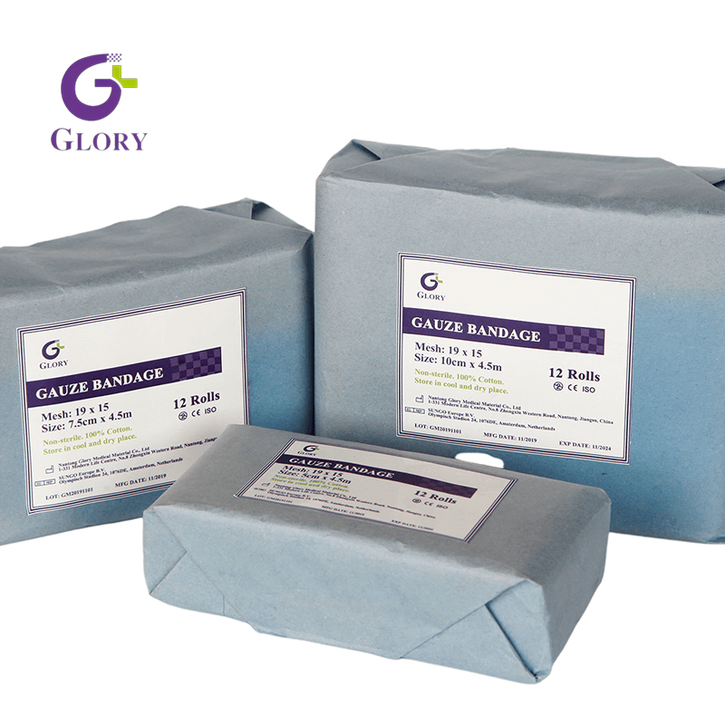
Traditional materials still hold a place in specific clinical scenarios. A soft gauze wound dressing serves primarily as a secondary layer. Clinicians historically used it for wet-to-dry mechanical debridement. Modern guidelines deprecate this practice due to tissue trauma. Gauze offers minor exudate management but requires frequent changes. It is highly permeable, allowing rapid moisture evaporation, which can lead to a desiccated wound bed if not monitored closely.
Gauze limitations are significant. It carries a high risk of adhering to the wound bed. Removing dried gauze causes secondary trauma and intense pain. It also sheds fibers into the wound, potentially delaying healing. When used as a primary dressing, it must be moistened with saline and changed before it dries completely to prevent mechanical stripping of new granulation tissue.
A paraffin gauze dressing addresses some adherence issues. It suits superficial wounds, minor burns, skin grafts, and donor sites. The soothing petrolatum contact layer prevents the secondary dressing from sticking to fragile tissue. It requires a secondary absorbent layer. If left in place too long without fluid exit pathways, it causes desiccation and tissue maceration. You must ensure the mesh size is appropriate for the exudate viscosity to prevent fluid trapping.
Advanced and Moisture-Retentive Dressings
Advanced materials actively manage the microenvironment. A foam wound dressing handles moderate to heavily exuding wounds. It is ideal for pressure ulcers, diabetic foot ulcers, and venous leg ulcers. The highly absorbent polyurethane or silicone structure provides thermal insulation. It offers moisture vapor transmission and cushioning for localized pressure offloading. The vertical wicking properties pull fluid away from the wound bed, locking it within the foam matrix to protect the periwound skin.
Foams are contraindicated for dry wounds or dry eschar. Their opaque nature prevents visual monitoring of the wound bed without physical removal. You must rely on exudate strike-through to gauge change frequency. Silicone-bordered foams offer atraumatic removal, making them excellent choices for patients with fragile, parchment-like skin.
Alginates and gelling fibers manage heavily exuding cavities and minor bleeding. Derived from seaweed, these sodium and calcium salts react with exudate. They form a cohesive hydrophilic gel. This gel promotes autolytic debridement and provides minor local hemostasis. You must apply a secondary dressing over alginates to secure them and manage excess moisture vapor.
Hydrogels and hydrocolloids serve distinct moisture needs. Hydrogels donate water to dry, necrotic tissue beds. They come in amorphous gels or sheet forms. Hydrocolloids suit low-to-moderate exuding chronic wounds. They provide an occlusive, self-adhesive barrier. This forms a gel-like mass over the wound to protect against external contaminants and promote autolytic debridement. Hydrocolloids often produce a characteristic odor upon removal as the gel breaks down, which you must differentiate from clinical infection.
Biological and Active Antimicrobial Dressings
Antimicrobial dressings utilize silver, cadexomer iodine, or PHMB. They target critically colonized or infected wounds with high bioburden. These dressings release antimicrobial agents directly into the wound bed. This controlled release reduces the bacterial load effectively. You should limit the use of active antimicrobials to a two-week challenge period. If the wound shows no improvement, reassess the bioburden and consider alternative interventions.
Collagen and active biological dressings jumpstart stalled chronic wounds. They are indicated for diabetic ulcers and pressure injuries showing no progress after four weeks of standard care. These dressings bind and inactivate destructive matrix metalloproteinases (MMPs). They simultaneously promote the deposition of new collagen fibers, providing a scaffold for cellular migration and tissue repair.
Patient-Centric and Operational Evaluation Dimensions
Anatomical Location and Conformability
The physical location of the wound dictates dressing requirements. Wounds on heels, sacrums, or joint creases demand high physical flexibility. The dressing must conform to complex contours without tenting or peeling. Self-adhesive properties and border requirements vary heavily based on anatomical placement. A sacral wound requires a specialized heart-shaped or butterfly-shaped dressing to prevent fecal contamination and withstand shearing forces during patient repositioning.
Rigid dressings fail on highly mobile joints. You must select conformable materials that move with the patient. This prevents premature detachment and maintains the protective barrier. Using liquid adhesives or secondary retention bandages helps secure dressings on difficult anatomical areas like the shoulder or the plantar surface of the foot.
Wear Time and Dressing Change Frequency
Wear time heavily impacts nursing staff bandwidth and clinical workflows. Frequent changes consume valuable clinical hours. You must establish the clinical threshold for leaving dressings undisturbed. Advanced dressings can remain in place for up to seven days, depending on exudate levels and manufacturer guidelines.
Undisturbed wear maintains optimal wound bed temperature. It supports uninterrupted cellular mitotic activity. Every dressing change drops the wound temperature, halting healing for several hours. Minimizing interventions accelerates the overall healing trajectory and reduces the risk of introducing exogenous bacteria into the wound bed.
Patient Quality of Life (QoL) and Compliance
Dressing choices directly impact patient mobility and physical comfort. You must assess their ability to bathe. Waterproof or water-resistant backings allow patients to maintain personal hygiene. This significantly boosts morale and compliance. Patients are more likely to adhere to a treatment plan if the dressing does not interfere with their daily activities.
Odor management is a key psychological factor. Malodorous wounds cause severe social isolation and distress. Modern wound dressing products incorporate charcoal or specialized barriers to neutralize odors. Improving patient well-being ensures better protocol adherence and reduces anxiety associated with chronic wound management.
Patient Sensitivities, Allergies, and Pain Management
Screening for patient allergies is mandatory. Common sensitivities include adhesives, acrylics, silver, iodine, or specific polymers. Applying an allergenic dressing causes severe contact dermatitis, worsening the wound condition. Always review the patient's medical history and perform a patch test if you suspect a material sensitivity.
Atraumatic removal technologies minimize pain. Soft silicone borders and medical adhesive removers prevent skin stripping. This is especially critical in fragile elderly or pediatric populations. Pain-free dressing changes build patient trust and compliance. Anticipating pain and utilizing appropriate analgesic strategies prior to dressing removal improves the overall patient experience.
Cost-to-Outcome Trade-Offs in Wound Care Procurement
Unit Cost vs. Total Cost of Healing
Selecting cheap materials often creates a financial fallacy. Basic gauze requires multiple daily changes. This consumes massive amounts of nursing time and auxiliary supplies. Higher-cost advanced dressings remain in place for days, drastically reducing labor requirements. The initial purchase price of a dressing represents only a fraction of the total intervention cost.
You must build a complete procurement evaluation model. Factor in labor costs, material disposal costs, and clinical complication management. Preventing secondary infections or maceration-induced wound expansion saves significant resources. The total cost of healing always outweighs the initial unit price. Investing in advanced moisture-retentive materials shortens the healing timeline and reduces hospital readmission rates.
Formulary Standardization vs. Specialized Inventory
Maintaining a streamlined facility formulary is a logistical challenge. You want cost-effective standardization. Clinicians need rapid access to specialized products for complex, non-healing wounds. Balancing these needs requires careful inventory management and clear clinical guidelines.
Establish clear clinical pathways for product escalation. Stock versatile, broad-spectrum dressings for general use. Maintain a controlled inventory of advanced biologicals and antimicrobials for specific, documented indications. Regular audits of dressing utilization help identify waste and ensure the formulary meets the actual clinical demands of the patient population.
Implementation Risks and Clinical Mitigation Strategies
Risk: Peri-Wound Maceration
Maceration occurs when excess fluid breaks down healthy periwound skin. This happens when you select a dressing with insufficient absorption capacity. Slow vertical wicking or a poor moisture vapor transmission rate also traps fluid against the skin. Macerated skin is highly susceptible to mechanical damage and bacterial penetration.
Apply a liquid skin protectant or barrier cream to the periwound area before dressing application.
Size the absorbent pad accurately so it does not overlap onto intact skin.
Step up to a high-capacity superabsorbent polymer dressing if exudate levels overwhelm standard foams.
Increase the frequency of dressing changes until exudate volume decreases.
Risk: Dressing Adherence and Trauma Upon Removal
Dry absorbent dressings stick to granulating tissue. Allowing a contact layer to desiccate within the wound bed causes severe removal trauma. This rips away newly formed cells and restarts the inflammatory phase. Repeated trauma delays healing indefinitely and increases patient pain.
Implement soft silicone contact layers to prevent adherence to the wound bed.
Utilize amorphous hydrogels to maintain a moist environment beneath absorbent secondary layers.
Moisten adhered dressings with sterile saline for several minutes prior to gentle removal.
Adjust clinical change frequency to ensure dressings do not dry out completely between assessments.
Risk: Undetected Infection
Prolonged wear time of thick, opaque dressings can mask clinical signs of infection. You might miss localized erythema, swelling, or purulent drainage until it becomes severe. Relying solely on scheduled change dates without interim assessments puts the patient at risk.
Establish strict clinical assessment protocols requiring daily visual inspection of the dressing margins.
Evaluate systemic signs of infection, such as fever, elevated white blood cell counts, or sudden changes in glycemic control.
Utilize dressings with transparent observation windows where appropriate to monitor the wound bed visually.
Educate patients and caregivers on the warning signs of infection and instruct them to report increased pain or odor immediately.
Conclusion
Optimal selection requires dynamic, phase-specific matching. You must align dressing capabilities with the physical and micro-environmental realities of the wound bed. Assess exudate volume and identify the tissue phase using the TIME framework. Evaluate infection risks carefully. Always factor in patient compliance, comfort, and the total cost of care when making your selection.
Conduct a comprehensive wound assessment using standardized clinical tools prior to selecting any product.
Consult with a certified wound care specialist for stalled, complex, or rapidly deteriorating wounds.
Review and update facility formularies regularly to optimize product availability based on current evidence.
Implement continuous hands-on training for nursing staff regarding advanced dressing application and removal techniques.
FAQ
Q: What is the most important factor when choosing a wound dressing?
A: Exudate management and maintaining balanced hydration are the primary drivers. You must match the dressing's absorption capacity to the wound's fluid output to prevent both desiccation and periwound maceration.
Q: How often should advanced wound dressings be changed?
A: Advanced dressings can often remain in place for 3 to 7 days, depending on exudate levels and manufacturer guidelines. Less frequent changes maintain optimal wound temperature and reduce tissue trauma.
Q: When should I use an antimicrobial dressing?
A: Use antimicrobial dressings when a wound is critically colonized or locally infected. They are not recommended for routine use on clean, granulating wounds due to potential cellular toxicity.
Q: Why is gauze no longer recommended for routine wound packing?
A: Gauze sheds fibers, adheres to the wound bed, and causes pain and trauma upon removal. It also requires frequent changes and fails to maintain an optimal moist healing environment.
Q: How do silicone dressings benefit the patient?
A: Soft silicone dressings provide atraumatic removal. They do not stick to the moist wound bed but adhere gently to dry periwound skin, minimizing pain and preventing skin stripping during changes.
